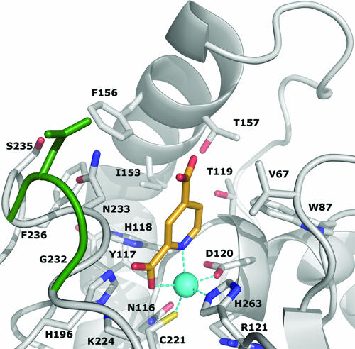
FIG. 4.
Active site of CphA-Zn(II) in complex with the pyridine-2,4-dicarboxylate inhibitor (carbon atoms colored in orange). The conformational change upon inhibitor binding is represented by superimposition of the wild-type Gly232 and Asn233 residues (green). The zinc ion is represented as a blue sphere. This figure was prepared using the program PYMOL.

