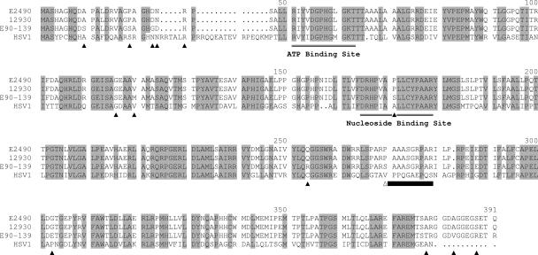
FIG. 2.
Alignment of BV and HSV-1 TK sequences. Amino acids constituting the ATP and the nucleoside binding sites are indicated, and all residues that are conserved between HSV-1 and all BV isolates are highlighted in gray. Residues that differ between the cynomolgous and rhesus BV sequences are indicated by black triangles under the sequences. The 10 amino acids (residues 270 to 279) deleted in the mutant recombinant BV TK referred to in the text are indicated by a black bar under the sequences.

