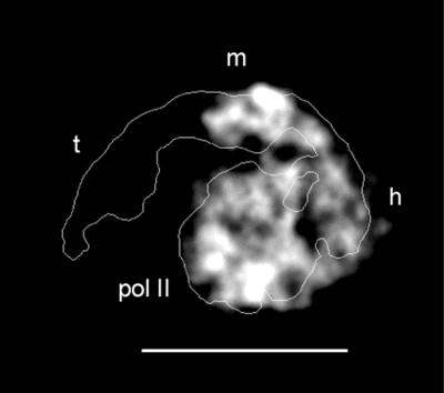
Figure 3.
Gray-scale projection map of a ΔSin4 mutant yeast holoenzyme (lacking subunits Sin4, Gal11, Med2, and Pgd1), calculated after alignment and averaging of ≈50 molecules preserved in uranyl acetate. (Bar = 200 Å.) An outline of the wild-type holoenzyme complex is shown for comparison (10). Mediator in the mutant holoenzyme appears wrapped around polymerase (pol II) in a extended conformation similar to that observed for the wild-type complex, but comparison of the structures reveals that the “tail” domain (labeled t in the wild-type holoenzyme outline) is missing, whereas the head (h) and middle (m) domains are present and interact with RNA polymerase as they do in the wild-type holoenzyme.

