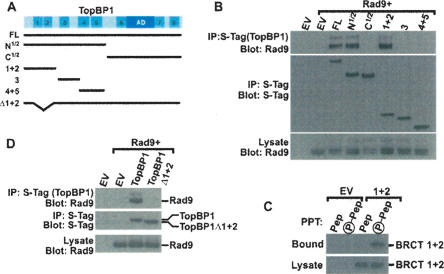
Figure 2.
TopBP1 BRCT domains 1 and 2 bind Rad9. (A) Schematic map of TopBP1 showing the BRCT domains, the AD, and the S-tagged constructs used in this study: full-length TopBP1 (FL); the N-terminal half of TopBP1 (N1/2); the C-terminal half of TopBP1 (C1/2); fragments encoding BRCT 1 and 2 (1 + 2), BRCT 3 (3), and BRCT 4 and 5 (4 + 5); and TopBP1 lacking BRCT 1 and 2 (Δ1 + 2). (B,D) HEK293 cells were transfected with empty vector (EV) or vectors encoding AU1-tagged Rad9 and the indicated S-tagged TopBP1 proteins shown in A. Lysates were precipitated with S-protein agarose beads. Bound proteins were sequentially immunoblotted for Rad9 (top) and S-tagged TopBP1 (middle). (Bottom) A portion of the lysate was immunoblotted with Rad9 to show equal expression. The multiple bands present in the Rad9 immunoblots are due to various forms of phosphorylated Rad9 (Volkmer and Karnitz 1999). (C) HEK293 cells were transfected with empty vector or a vector expressing S-tagged TopBP1 BRCT 1 and 2 domains (1 + 2). (Top) Lysates were precipitated with nonphosphorylated (Pep) or phosphorylated (P-Pep) Ser387 Rad9 peptide covalently linked to beads. (Bottom) A portion of the lysate was immunoblotted to demonstrate equal expression of the BRCT 1 + 2 domains.

