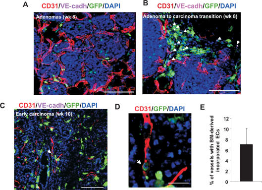
Figure 4.
Contribution of BM-derived EPCs in spontaneous breast tumors. (A) Primary adenoma lesions in mammary gland sections from a PyMT mouse at 8 wk of age. Pre-existing CD31+ vessels are observed surrounding the adenomas. Bar, 100 μm. (B) Recruitment of BM-derived GFP+ VE-cadherin+ EPCs (arrows) at the periphery of the avascular adenoma–carcinoma progression is shown. Bar, 100 μm. (C) An early carcinoma showing recruited CD31+ mature vessels in the tumor mass (10 wk of age). Arrow depicts incorporated BM-derived ECs in a vessel. Bar, 100 μm. (D) High-resolution image of a representative blood vessel (box in C) showing an incorporated mature BM-derived GFP+ CD31+-coexpressing cell (arrow). Bar, 20 μm. DAPI was used to stain the nucleus of all cells. (E) Quantification of vessels in breast tumors (10 wk old) with incorporated BM-derived ECs (GFP+ CD31+ VE-cadherin+). A minimum of 250 vessels were counted from nonsequential sections from four animals, and Z-stacks were evaluated for each section. Error bars represent standard deviations.

