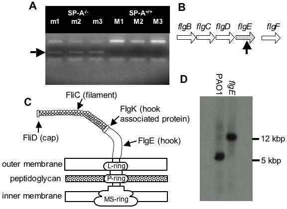
Figure 1. The Flagellar Mutant flgE Is Preferentially Cleared from the Lungs of SP-A+/+ Mice.

(A) Pools of 72 uniquely-tagged mutants were intranasally inoculated into three SP-A+/+ (M1, M2, M3) and three SP-A−/− (m1, m2, m3) mice. Mouse lungs were harvested, homogenized and plated. Bacteria colonies were collected for genomic DNA extraction. PCR-amplification of tags was performed to screen for the presence or absence of each of the 72 mutants. The band indicative of the flgE mutant was present in the SP-A−/− pool, but absent in the SP-A+/+ pool. (B) Genetic organization of the flg operon in P. aeruginosa. The horizontal arrows (flgB, flgC, flgD, flgE, flgF) represent transcriptional directions of the genes coding for flagella proteins. The vertical black arrow indicates the approximate insertion site within the mutated open reading frame of flgE. (C) A schematic representation of flagellar structure. FliC is flagellar filament protein, FliD is flagellar cap protein, FlgE is flagellar hook protein, and FlgK is flagellar hook associated protein. (D) Restriction fragment length polymorphism analysis between wild-type PA01 and flgE mutant indicates that the flgE mutation was caused by transposon insertion into the cloned gene.
