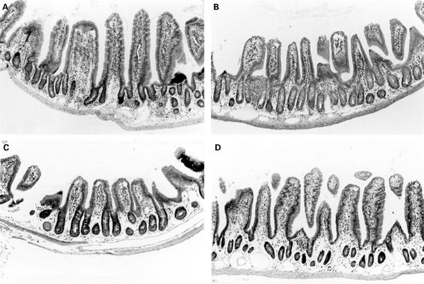
Figure 6 .
: Occurrence of intestinal lesions in B6 nu/nu mice inoculated with MAIDS spleen cells. A: thickened intestinal wall in B6 nu/nu mice inoculated with MAIDS spleen cells. Cellular infiltration is localised in the lamina propria and submucosa of the small intestine. Epithelial cell hyperplasia is also observed but neither erosion nor ulcer is observed; B-D: controls; small intestines of B6 nu/nu mice inoculated with B6 spleen cells (B), untreated B6 nu/nu mice (C), and B6 nu/nu mice infected with LP-BM5 (D). (Haematoxylin and eosin; original magnification: ×120.)

