Abstract
Background—Recent clinical studies suggest that ischaemic colitis is caused by a microcirculatory disturbance that involves thrombosis of the colon. Aim—To establish a new model of photochemically induced ischaemic colitis in rats. Methods—Thirty male Wistar rats were anaesthetised with amobarbital, the femoral veins were cannulated and laparotomies were performed. The serosal surface of the proximal colon was irradiated by using a krypton laser (wavelength 568 nm, 20 mW) for four minutes. An intravenous infusion of a photosensitising dye, rose bengal (20 mg/kg body weight), was administered over 90 seconds, beginning at the start of irradiation. Rats were killed immediately (n=4), 12 hours (n=2), 24 hours (n=10), three days (n=4), seven days (n=4), 14 days (n=2), or 28 days (n=2) after irradiation. Two control rats received laser irradiation without dye infusion. Specimens of the irradiated sites were examined by using histopathology. Results—Localised ulcers of the colon were present in rats killed at 12 hours, 24 hours, three days, and seven days after irradiation. Microscopy findings were consistent with the features of human ischaemic colitis. Reproducible ulcerative lesions were produced by photothrombosis of microvessels in the colon. Conclusion—This model may be useful for further investigation of the pathophysiology of ischaemic colitis.
Keywords: ischaemic colitis; photothrombosis; rose bengal
Full Text
The Full Text of this article is available as a PDF (160.2 KB).
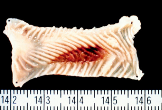
Figure 1 .
: Macroscopic findings showing a colonic intraluminal lesion in a laser-irradiated rat at 24 hours. Localised shallow open ulcers with irregular margins are evident.
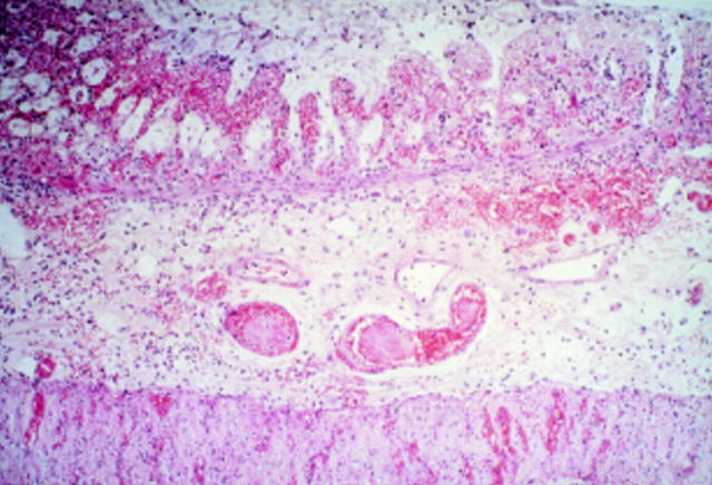
Figure 2 .
: Histological findings of a photochemically induced ulcer of the colon. The ulcer was restricted to the mucosa or submucosa, which showed fresh mucosal haemorrhage, degeneration and necrosis of the glandular epithelium with a "ghost-like" appearance, and notable submucosal thickening caused by prominent oedema, associated with congestion and neutrophilic infiltration.
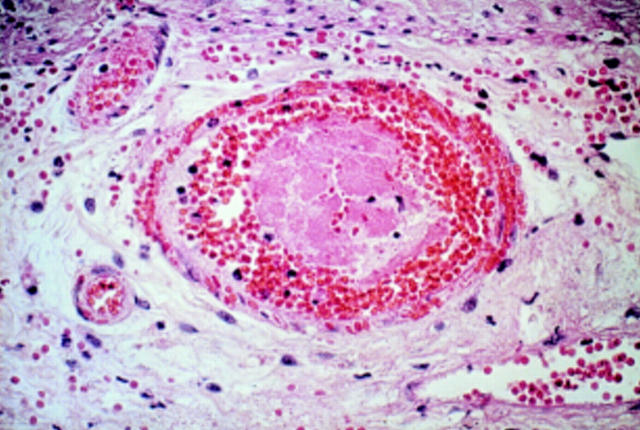
Figure 3 .
: Microscopic findings of an ulcer in the laser-irradiated colon of a rat. The microvessel of the submucosal layer was filled with photochemically induced thrombi.
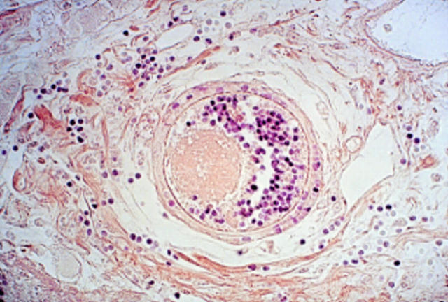
Figure 4 .
: The photothrombi were not stained by PTAH, indicating the absence of a fibrin component.
Selected References
These references are in PubMed. This may not be the complete list of references from this article.
- BOLEY S. J., SCHWARTZ S., LASH J., STERNHILL V. Reversible vascular occlusion of the colon. Surg Gynecol Obstet. 1963 Jan;116:53–60. [PubMed] [Google Scholar]
- Boley S. J., Krieger H., Schultz L., Robinson K., Siew F. P., Allen A. C., Schwartz S. Experimental aspects of peripheral vascular occlusion of the intestine. Surg Gynecol Obstet. 1965 Oct;121(4):789–794. [PubMed] [Google Scholar]
- Iida M., Matsui T., Fuchigami T., Iwashita A., Yao T., Fujishima M. Clinical features of nongangrenous ischemic colitis. A comparison of stricturing and transient forms. J Clin Gastroenterol. 1986 Dec;8(6):635–639. doi: 10.1097/00004836-198612000-00009. [DOI] [PubMed] [Google Scholar]
- Iida M., Matsui T., Fuchigami T., Iwashita A., Yao T., Fujishima M. Ischemic colitis: serial changes in double-contrast barium enema examination. Radiology. 1986 May;159(2):337–341. doi: 10.1148/radiology.159.2.3961164. [DOI] [PubMed] [Google Scholar]
- Marston A., Pheils M. T., Thomas M. L., Morson B. C. Ischaemic colitis. Gut. 1966 Feb;7(1):1–15. doi: 10.1136/gut.7.1.1. [DOI] [PMC free article] [PubMed] [Google Scholar]
- Matsumoto T., Iida M., Nakamura S., Hizawa K., Kuroki F., Fujishima M. An animal model of longitudinal ulcers in the small intestine induced by intracolonically administered indomethacin in rats. Gastroenterol Jpn. 1993 Feb;28(1):10–17. doi: 10.1007/BF02774998. [DOI] [PubMed] [Google Scholar]
- Mishima Y., Horie Y. Experimental studies of ischemic enterocolitis. World J Surg. 1980 Sep;4(5):601–605. doi: 10.1007/BF02401641. [DOI] [PubMed] [Google Scholar]
- Ruf W., Suehiro G. T., Suehiro A., Pressler V., McNamara J. J. Intestinal blood flow at various intraluminal pressures in the piglet with closed abdomen. Ann Surg. 1980 Feb;191(2):157–163. doi: 10.1097/00000658-198002000-00005. [DOI] [PMC free article] [PubMed] [Google Scholar]
- Watson B. D., Dietrich W. D., Busto R., Wachtel M. S., Ginsberg M. D. Induction of reproducible brain infarction by photochemically initiated thrombosis. Ann Neurol. 1985 May;17(5):497–504. doi: 10.1002/ana.410170513. [DOI] [PubMed] [Google Scholar]
- Yao H., Ibayashi S., Sugimori H., Fujii K., Fujishima M. Simplified model of krypton laser-induced thrombotic distal middle cerebral artery occlusion in spontaneously hypertensive rats. Stroke. 1996 Feb;27(2):333–336. doi: 10.1161/01.str.27.2.333. [DOI] [PubMed] [Google Scholar]


