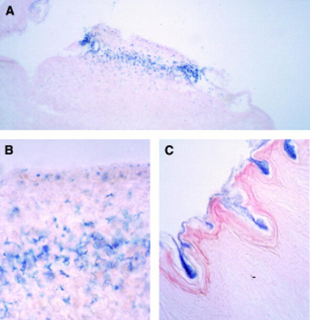
Figure 2 .
: Microscopic view of sections from transduced oesophageal epithelium using the double balloon catheter system. (A) Representative area positively stained for β-galactosidase activity (original magnification ×50); (B) an oesophageal section three days after luminal gene transfer, indicating transfected epithelial layers (original magnification ×250); (C) an oesophageal section with an intact keratin layer which acts as a barrier for gene transfer (original magnification ×100). Sections were counterstained with eosin.

