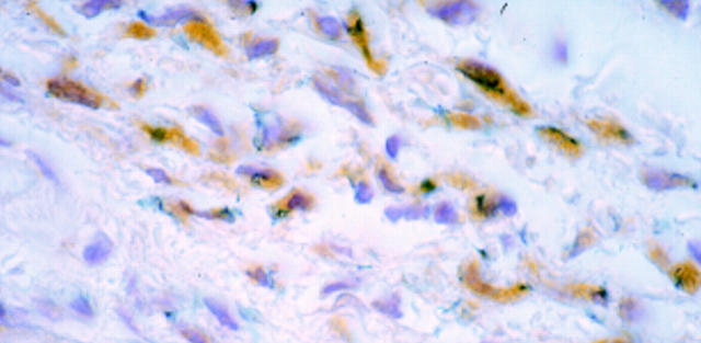
Figure 4 .
: Microscopic view of sections from oesophagus transduced by injection. Sections were incubated with a monoclonal anti-β-galactosidase antibody followed by a secondary biotin conjugated rabbit antimouse antibody and streptavidin-peroxidase complexes with DAB. β-Galactosidase activity is presented as a blue precipitate, whereas detection by the anti-β-galactosidase antibody is displayed as brown precipitate throughout the cytoplasm. The section was counterstained with haematoxylin. Original magnification ×250.

