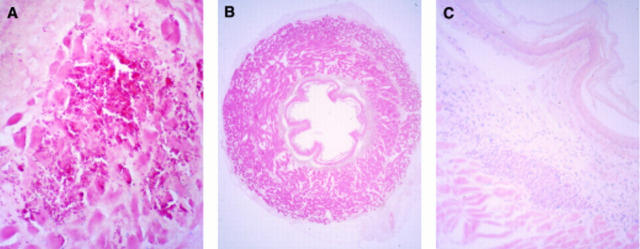
Figure 5 .
: Microscopic view of a section from the oesophagus injected with 200 µl DNA-liposomes. Three days following gene transfer, signs of inflammation, necrosis, and bleeding were visible at the area where the injection took place. Sections were fixed in 4% paraformaldehyde and stained with eosin (A) (original magnification ×50). Twenty eight days after gene transfer no signs of trauma, necrosis, or major inflammation were visible (B) (original magnification ×6.25). In these areas a local increase in small lymphoid aggregates remained in the submucosa (C) (original magnification ×50). Sections were counterstained with eosin (A) or with eosin in combination with haematoxylin (B and C).

