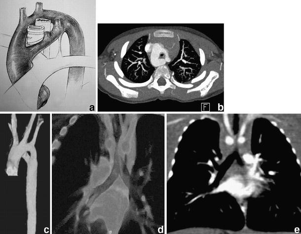
Fig. 11.

Complete vascular ring around the trachea and the oesophagus formed by a double aortic arch. a Diagrammatic representation of the abnormality. b Corresponding contrast-enhanced axial image at the level of the double aortic arch, the right part being the dominant. c 3-D VR image of the double aortic arch from a left anterior oblique view. Volume slab (d) and coronal MPR (e) additionally demonstrate the resulting significant narrowing of the trachea
