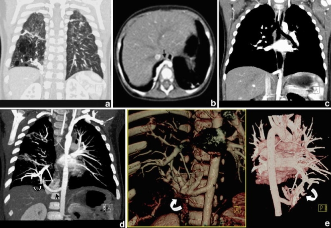
Fig. 3.

Combiscan in an 11-month-old child with a congenital pulmonary airway malformation in the right lower lobe. a Coronal MPR in lung window setting shows the air-filled cystic component of the lesion. b, c Axial CT slice (b) and coronal MPR (c) showing a large arterial feeder (arrow). This is, however, visualized and appreciated better with an oblique MIP along the axis of the vessel (d) and with coronal anterior and posterior 3-D VR (e). These images additionally demonstrate the pulmonary venous drainage of the lesion, i.e. intralobar sequestration (curved arrows)
