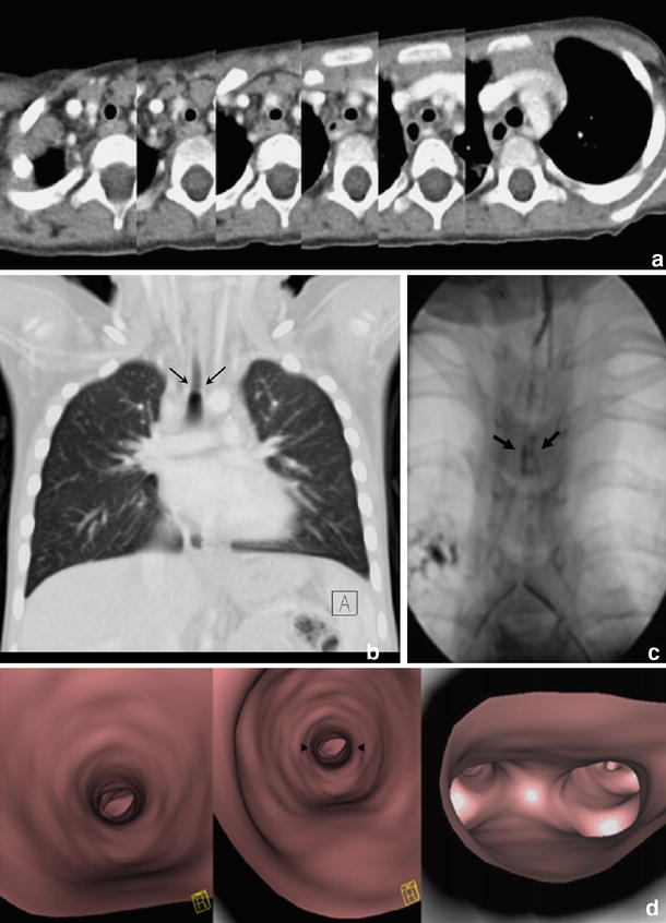
Fig. 4.

Congenital tracheal stenosis in a 3-year-old girl with trisomy 21. a Series of axial slices demonstrating stenosis in the central third of the trachea. b This is more sensitively demonstrated on coronal MPR, which correlates well with the bronchographic appearances(c) (arrows). d Virtual bronchoscopy also demonstrates mild stenosis of the supracarinal portion of the trachea (arrowheads)
