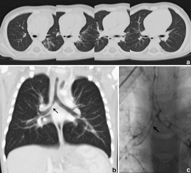
Fig. 5.

Chest CT (1.5 mm collimation following intravenous injection of contrast medium) in 10-month-old boy with recurrent wheezing due to congenital tracheal stenosis treated previously with tracheoplasty. There is mild stenosis of the trachea and the origin of the right main bronchus, which is appreciated with difficulty on the axial scans(a), but is nicely shown in coronal MPR (arrow) (b). Virtual bronchoscopy in this case was unnecessary. Bronchography (c) and bronchoscopy confirmed stenosis in the right main bronchus (arrow) that was caused by granulation tissue
