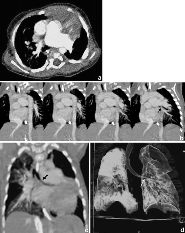
Fig. 6.

Cardiac CT (0.75 collimation after intravenous injection of contrast medium) in a 6-month-old girl with absent pulmonary valve syndrome. a Axial contrast-enhanced CT shows massive dilatation of the main and the left pulmonary arteries with possible compression of the left main bronchus. This is also appreciated on serial oblique MIPs (b), but becomes more apparent on the oblique MIP along the axis of the left main bronchus (c) (black arrow) and on the 3-D VR (d) (white arrow)
