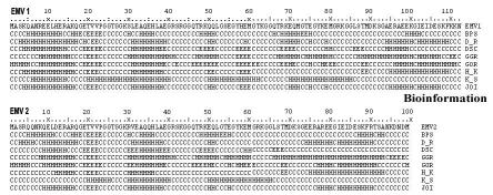
Figure 2.
Secondary structure analysis of the predicted EMV proteins performed with PELE program available on the SDSC Biology Workbench (http://workbench.sdsc.edu [13]). Seven different structure predictions are shown, with the most likely structural feature at each residue indicated by H (α-helices), E (β-sheets) or C (random coils). The programs used are denoted BPS, D_R, DSC, GGR, GOR, H_K and K_S. The “winner-takes-all” joint prediction was given by the JOI program.

