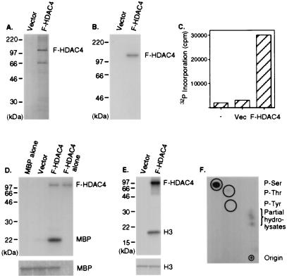
Figure 1.
Serine/threonine protein kinases associate with F-HDAC4 complexes and phosphorylate F-HDAC4. (A and B) Anti-FLAG immunoprecipitates were resolved by SDS/PAGE. Commassie blue staining (A) and Western blot analysis (B) with anti-FLAG antibody after blotting proteins onto a nitrocellulose filter. Vec, vector. (C–E) The presence of protein kinase activity was assayed by using the following substrates: synthetic peptide KKALRRQETVDAL (C), MBP (D), and histone H3 in the presence of [γ-32P]ATP (E) . D and E show results from autoradiography (Upper) and Commassie blue stain (Lower). (F) Two-dimensional-electrophoresis analysis of acid-hydrolyzed phosphorylated F-HDAC4 from the gel shown in E. The positions of the origin, cold phosphoamino acid standards, and partially hydrolyzed peptide are shown. P-Ser, phosphoserine; P-Thr, phosphothreonine; P-Tyr, phosphotyrosine.

