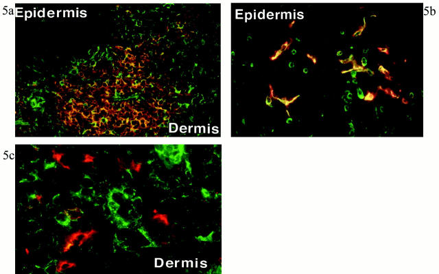
Figure 5.
Double-immunofluorescence staining for CCR4 and cell markers in inflamed skin. Results shown in a, b, and c are representative figures of five experiments of this combination with inflamed skin. a: Numerous CCR4+ cells (green) and CD4+ cells (orange) are present in the T cell-DC cluster of dermis. CCR4+CD4+ cells (bright yellow) constitute approximately one-third of CD4+ cells. A part of CD4+ T cells in the epidermis also expressed CCR4. b: Double immunofluorescence for CCR4 (green) and CD1a (red) in the epidermis. Approximately half of epidermal LCs express CCR4 (yellow). c: Double immunofluorescence for CCR4 (green) and CD83 (red) in the inflamed dermis. CCR4+ cells and CD83+ cells are different. Focal yellow color may represent overlapping of cell membranes in thick frozen section. Original magnifications, ×100 (a), ×300 (b), ×320 (c).

