Figure 1.
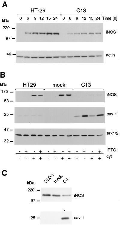
Levels of iNOS protein were reduced on ectopic expression of cav-1 in HT29 and DLD1 cells. iNOS and cav-1 protein levels were investigated by Western blot analysis. (A) The kinetics of cytokine-induced iNOS protein expression in HT29 and cav-1-expressing C13 cells is shown. Reduced levels of iNOS expression were apparent as early as 6 h after cytokine stimulation and remained low throughout the experiment up to 24 h after induction. Actin was used as control for protein loading. (B) Analysis of parental, mock-, and cav-1-transfected (C13) HT29 cells in the presence (+) or absence (−) of cytokines (cyt) and/or IPTG (150 μM). Extracellular signal-regulated kinase 1/2 (erk1/2) was used as control for protein loading in each lane. (C) iNOS and cav-1 protein levels were determined in DLD1-, mock-, and cav-1-transfected C4 cells after stimulation of the cells with cytokines. As above for HT29 cells, iNOS protein levels were substantially lower in cells ectopically expressing cav-1. Migration positions of molecular-mass marker proteins are indicated to the left of the individual panels.

