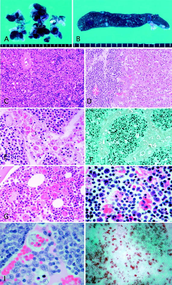
Figure 1.

A: A marked swelling of dark purple lymph nodes with severe hemophagocytosis. B: Cross-section of the enlarged spleen showing small white nodular lesions. C: Diffuse infiltration of atypical large lymphoid cells in the spleen. D: Periportal infiltration of atypical large lymphoid cells and marked central necrosis of the hepatic lobule. E: The lymph node showing diffuse infiltration of atypical lymphoid cells and marked erythrophagocytosis in the sinus. F: Atypical lymphoid cells with EBER1 expression diffusely infiltrated the parenchyma of the lymph node with severe sinus hemophagocytosis. G: Hemophagocytosis in the bone marrow. H Infiltrated atypical lymphocytes and hemophagocytic cells with multiple ingested cell debris, lymphocytes, and erythrocytes observed in the thymus. I: High-power view of the lymph node showing atypical large lymphoid cell infiltration in both sinus and medulla. J: Marked infiltration of rabbit CD5-positive lymphoid cells in the lymph node. Original magnifications: ×150 (hematoxylin and eosin; C and D), ×300 (hematoxylin and eosin; E and G), ×750 (hematoxylin and eosin; H and I), ×150 (EBER1 in situ hybridization; F), and ×200 (rabbit CD5; J).
