
Figure 8.
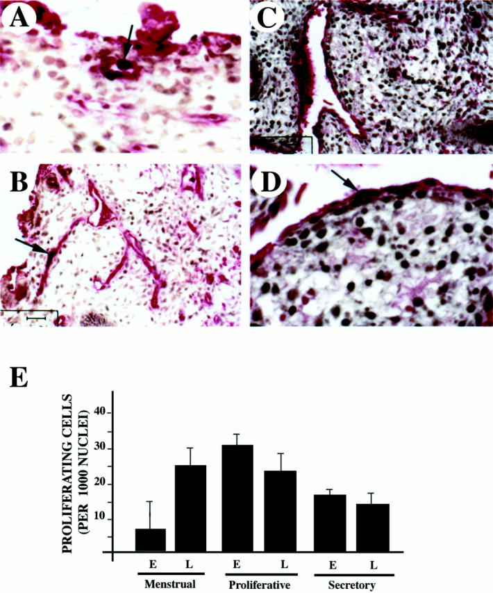
Proliferation of endothelial cells is initiated during the late menstrual phase. Capillaries were detected with anti-von Willebrand factor (in red) using an alkaline-phosphatase reaction and proliferating cells were detected with Ki67 antigen (in black) (light brown staining is background) in sections of menstrual (A and B) and early proliferative endometrium (C and D). Arrows indicate proliferating endothelial cells. E: Quantification of proliferating endothelial cells during early (E) and late (L) menstrual, proliferating and secretory phases. Six independent samples from each phase were used in the assessment. Numbers correspond to proliferating cells per 1,000 endothelial nuclei.