
Figure 3.
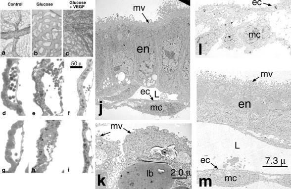
Hyperglycemia-induced disorganization of endothelial cells and adjacent mural cells is rescued by treatment with exogenous VEGF-A165. Low power representative micrograph of PECAM-1 stained 9.5 dpc yolk sacs harvested from normoglycemic (a), hyperglycemic (b), and hyperglycemic + exogenous VEGF-A165 cultures (c). Note the lack of arborization and ectatic vessels in the hyperglycemia-exposed conceptuses (b) and the rescue of an arborizing phenotype in the in the hyperglycemia-exposed conceptuses treated with exogenous VEGF-A165 (c). d–i: High power micrographs illustrating the perturbation of the intimate endothelial cell-mural cell interactions following hyperglycemic insult at 8.5 (compare normoglycemic, d, with e) and 9.5 dpc (compare normoglycemic, g, with h). f and i: Illustrations of the rescue of intimate endothelial cell-mural cell interactions after addition of exogenous rVEGF-A165 to the cultures (compare d with f and g with i). Scale bar, 50 μm (d–i). j–m: Representative transmission electron micrographs of control (j), hyperglycemic (k and l), and hyperglycemic 9.5 dpc conceptuses treated with VEGF-A165 (m). j: Illustratation of a yolk sac comprised of polarized endodermal cells (en) with apical microvilli (mv) and a microvascular lumen (L), lined by flattened endothelial cells (ec) intimately invested by mesothelial cells (mc). In contrast, k illustrates a hyperglycemic yolk sac comprised of endodermal cells with blunted microvilli and large lysosomal bodies (lb). l: Illustratation of the yolk sac vasculature, which is comprised of plump endothelial cells (ec) and mesothelial cells (mc) that have lost their intimate associations with the endothelium. m: A hyperglycemic yolk sac treated with exogenous rVEGF-A165. Its endodermal cells (en) are polarized with apical microvilli (mv). Its vasculature is again noted to be comprised of flattened endothelial cells (ec) intimately invested by mesothelial cells (mc). Scale bars, 7.3 μm (j, l, and m) and 2.0 μm (k).