
Figure 1.
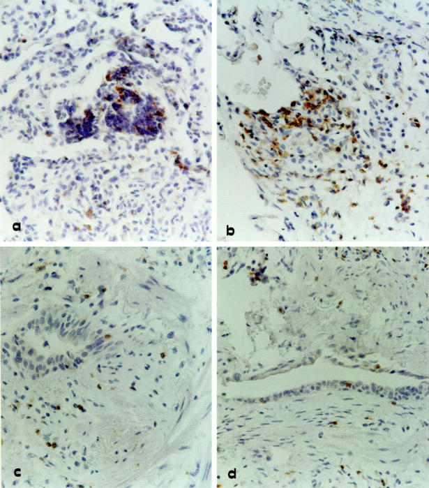
Immunohistochemistry for CXCR3 expression by lung T cells infiltrating transbronchial biopsy of four representative allograft recipients. a and b: Two allograft recipients with a rejection episode classified as A2 and A3 grades, respectively. c and d: Two representative lung allograft recipients with active bronchiolitis obliterans. Strongly stained CXCR3+ lymphocytes can be demonstrated not only in the perivascular space but also in the alveolar septa and air spaces of patients with acute cellular rejection (a and b). In patients with active obliterative bronchiolitis (c and d) subepithelial fibrosis can be observed in the bronchiole and is associated with a moderate infiltrate of CXCR3 stained lymphocytes. Original magnification, ×400.