
Figure 4.
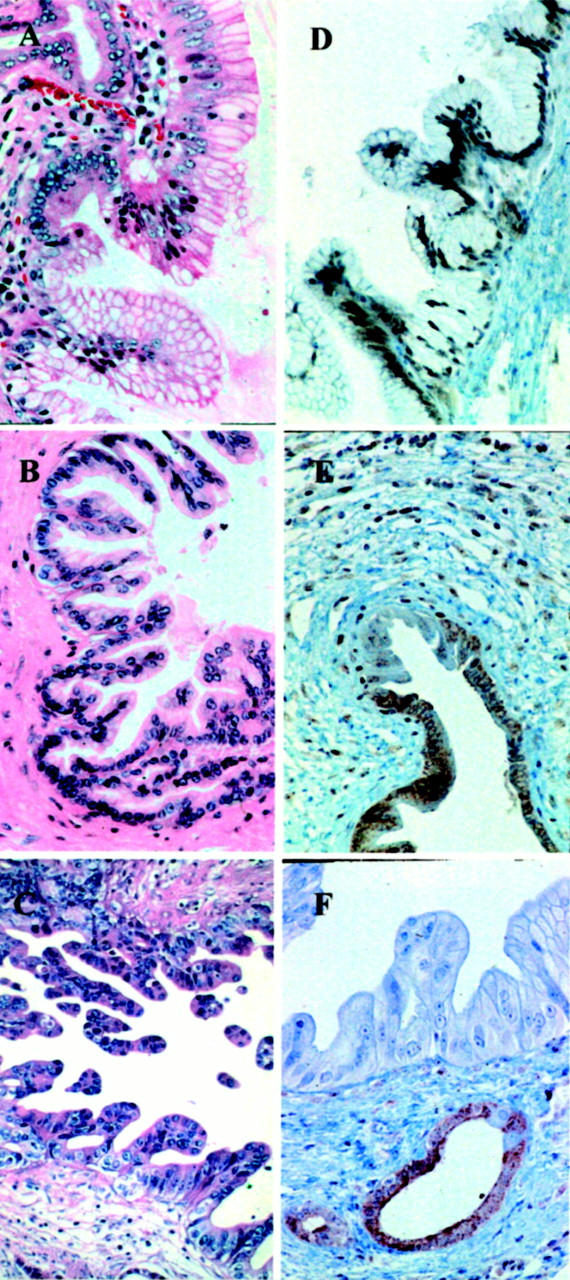
Examples of various grades of duct lesions associated with pancreatic carcinoma. A: Papillary duct lesion, grade PanIN-2 with low-grade dysplasia: slight nuclear enlargement and nuclear crowding. H&E; original magnification, ×250. B: Papillary duct lesion, grade PanIN-2 with moderate-grade dysplasia: moderate nuclear enlargement, nuclear hyperchromasia and loss of polarity. H&E; original magnification, ×250. C: Papillary duct lesion, grade PanIN-3: loss of polarity and structure and increasing branching of the papillae. H&E; original magnification, ×250. D–F: Immunohistochemical staining for Dpc4. D: Duct lesion, mainly PanIN-2 with low-grade dysplasia and partially PanIN-1A, both with positive staining for Dpc4 protein. Original magnification, ×125. E: Duct lesion, grade PanIN-2 with moderate-type dysplasia with nuclear Dpc4 staining, and a few negative nuclei in the adjacent intraductal carcinoma. Original magnification, ×125. F: Duct lesion, mainly grade PanIN-3 without Dpc4 protein expression, but retained Dpc4 expression in adjacent hyperplastic epithelium. Original magnification, ×250.