
Figure 3.
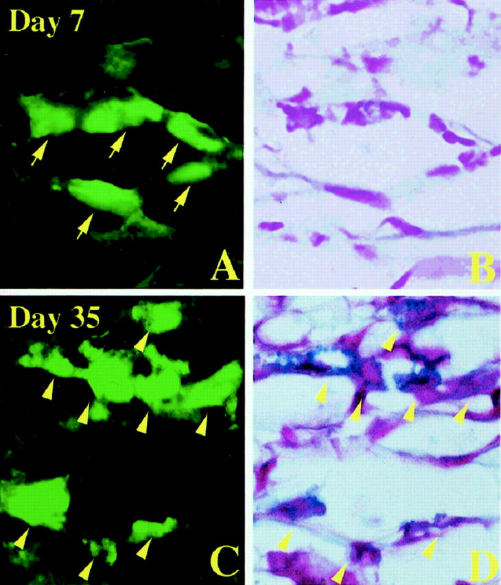
Granulated and degranulated mast cells after injection of 500 PMCs of WBB6F1-c-kit+/c-kit+; GFP+/GFP− mice into the stomach of WBB6F1-c-kitW/c-kitWv; GFP−/GFP− mice. A: GFP+ cells in the muscularis propria on day 7 after the injection. B: Same section of A stained with AB and nuclear fast red. GFP+ cells shown by arrows in A were not stained with AB. C: GFP+ cells in the muscularis propria on day 35 after the injection. D: Same section of C stained with AB and nuclear fast red. GFP+ cells shown by arrowheads in C were also stained with Alcian blue. Original magnification, ×1000.