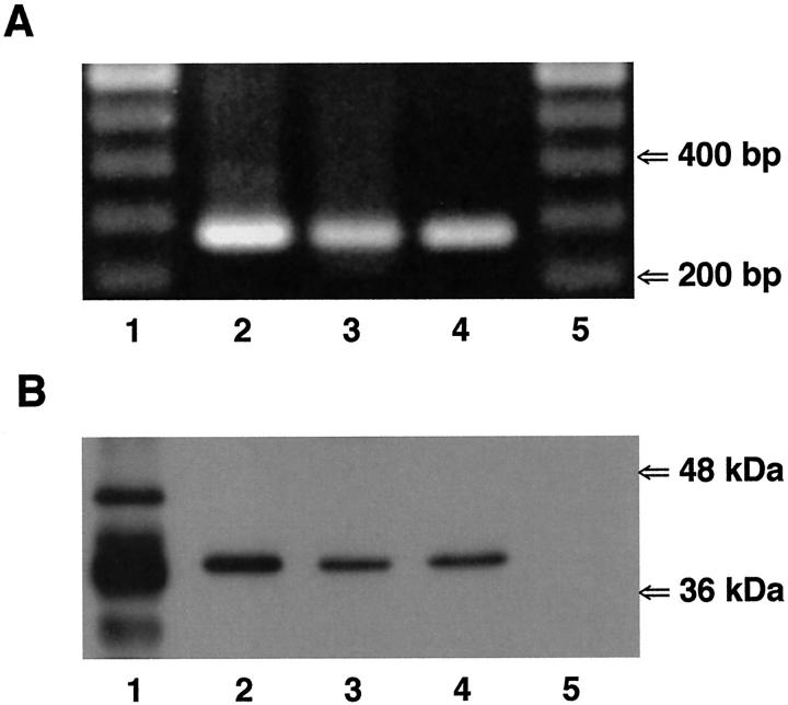
Figure 3.
Expression of Cx43 in non-CF and CF airway cells. A: Reverse transcriptase-polymerase chain reaction was performed on mRNA isolated from Beas2B (lane 2), CF15 (lane 3), and IB3-1 (lane 4) cells using primer pairs specific for human Cx43. Amplification products of the expected sizes for Cx43 (285 bp) were detected in all non-CF and CF cell lines. Molecular markers are shown in lanes 1 and 5. B: Western blot analysis of Cx43 expression in non-CF and CF airway cells. The anti-Cx43 antibody revealed one band at ∼41 kd in cytosolic protein extracts from Beas2B (lane 2), CF15 (lane 3), and IB3-1 (lane 4) cells. Lane 1 corresponds to rat atrium samples that were used as positive controls. Several bands at 41 to 46 kd could be detected, corresponding to various phosphorylated forms of Cx43. The lower band likely corresponds to a degradation product of Cx43. Lane 5 corresponds to SKHep1 cells, which do not express Cx43, that were used as negative controls.

