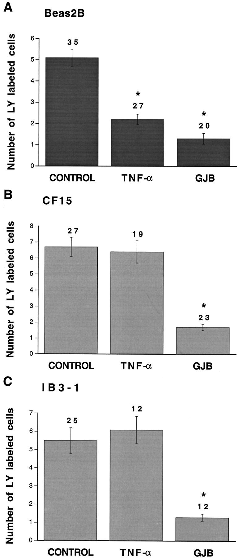
Figure 6.

Quantitative evaluation of dye coupling in non-CF and CF airway cells exposed to TNF-α. Under control conditions, transfer of lucifer yellow (LY) was detected in Beas2B (A), CF15 (B), and IB3-1 (C) cells. Whereas TNF-α significantly decreased dye transfer in non-CF Beas2B cells, the pro-inflammatory mediator had no effect on intercellular communication between CF (CF15 and IB3-1) cells. In all cells lines, gap junction channel blockers (GJB) inhibited dye coupling. Asterisks indicate differences at P < 0.002 levels.
