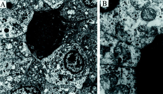
Figure 9.
Electron microscopy of tumor from patient 1. A: Low-power electron micrograph shows tumor cells surrounding a pool of basement membrane material. The cytoplasm of the tumor cells contains prominent mitochondria, scattered membrane-bound granules, and varying amounts of glycogen (uranyl acetate and lead citrate stain; original magnification, ×3000). B: Higher power micrograph shows the multilamellar appearance of the basement membrane material, and an intercellular junction (arrow) (uranyl acetate and lead citrate stain; original magnification, ×13,000).

