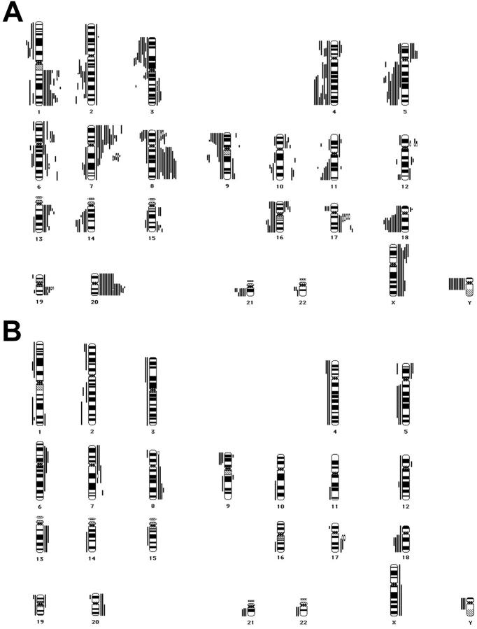
Figure 1.
A: Chromosomal ideograms showing the summary of DNA copy number changes, detected by CGH in 20 gastric cardia adenocarcinomas. Losses are displayed (as bars) on the left of the ideogram, gains are shown on the right. Frequent loss is seen on 1p, 2q, 3p, 4pq, 5q, 8p, 9pq, 11q, 14q, 16q, 18q, 21q, and Y. Frequent gain is detected at 1q, 5p, 6pq, 7pq, 8pq, 13q, 17q, 19q, 20pq, and Xpq. High level amplification (HLA; marked by an open bar) was frequently detected on 7q21, 8p22, 12p11.2, 17q12-q21, and 19q13.1-q13.2. B: Chromosomal ideograms showing the summary of DNA copy number changes, detected by CGH in 10 preneoplastic lesions (4 metaplasias, 1 low-grade dysplasia, 5 high-grade dysplasias). Frequent loss can be discriminated on 2q, 4p, 5q, 9p, 18q, 21q, and Y; frequent gain is seen on 6p, 7q, 8q, 13q, 17q, and 20q. HLAs were found only in high-grade dysplasia on 7q21, 8p22, and 17q12-q21.

