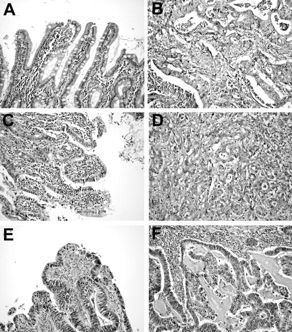
Figure 2.

Photomicrographs illustrating pairs of preneoplastic lesions (A, C, and E) and adenocarcinoma (B, D, and F) of the gastric cardia. Case 3 showed intestinal metaplasia (A) adjacent to poorly differentiated adenocarcinoma (B). Precursor and cancer shared gain of the X chromosome, whereas in this metaplastic tissue also loss on 18q was disclosed by CGH. Case 6 displayed high-grade dysplasia (C) and poorly differentiated adenocarcinoma (D). Many alterations (12 of 17) detected by CGH in the dysplasia were also discerned in the invasive cancer. Case 7 demonstrated HGD (E) and moderately differentiated adenocarcinoma (F). CGH revealed a basically similar pattern of chromosomal aberrations, despite the clear differences in histomorphology of the two lesions, ie, a non-invasive histology in HGD versus the invasive growth pattern of the adenocarcinoma.
