
Figure 3.
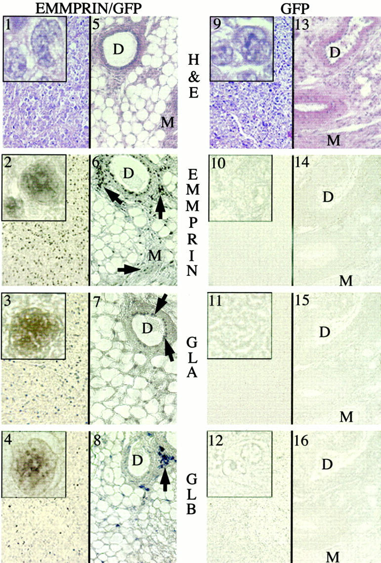
In situ hybridization of primary tumors from mice injected with EMMPRIN/GFP and GFP-transfected MDA-MB-436 breast cancer cells. Serial sections from tumor tissue (panels 1–4, and 9–12) and surrounding nonmalignant tissue (panels 5–8 and 13–16) were examined. Panels 1 and 9 represent H&E staining of cancer tissues from EMMPRIN/GFP and GFP tumors, respectively; panels 5 and 13 represent H&E staining of non malignant mammary tissues (tumor cells not identified) adjacent to the primary EMMPRIN/GFP and GFP tumors, respectively. Cells in the primary tumor mass from mice injected with EMMPRIN/GFP-transfected cells revealed widely distributed, specific staining with EMMPRIN, gelatinase A (GLA), and gelatinase B (GLB) antisense riboprobes (panels 2–4, respectively). Minimal cell staining for EMMPRIN, gelatinase A, and gelatinase B was seen in cancer cells from GFP-transfected MDA-MB-436 cells (panels 10–12). Nonmalignant tissues adjacent to the primary tumors from EMMPRIN/GFP mice demonstrated focal staining for EMMPRIN, gelatinase A, and gelatinase B in mammary ducts (D) and myocytes (M) (panels 6–8: arrows identify EMMPRIN-expressing cells). Nonmalignant tissue from GFP mice revealed no discernable staining for EMMPRIN, gelatinase A, or gelatinase B (panels 14–16). The insets in panels 1–4 display higher magnifications of mRNA-stained tumor cells; prominent nuclear staining is noted. Comparable cells (but minimally stained) in the GFP-transfected tumors are demonstrated in panels 10–12.