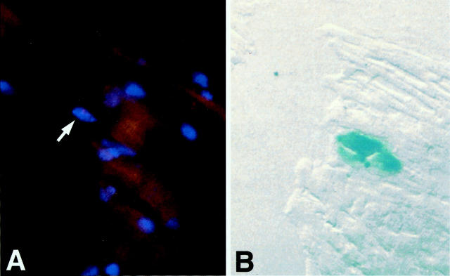
Figure 3.
Fluorescence in situ hybridization. The pink Y-chromosome (arrow) and the blue X-gal product are demonstrated in adjoining 8-μm serial sections of the same cell. A: Rhodamine staining of the Y-chromosome probed with a rat Y-chromosome-specific repetitive DNA sequence (arrow). B: Adjoining section of the same myocyte, the blue color is from the X-gal reaction. Superimposition of both signals is affected by an 8-μm shift in register of the two serial sections.

