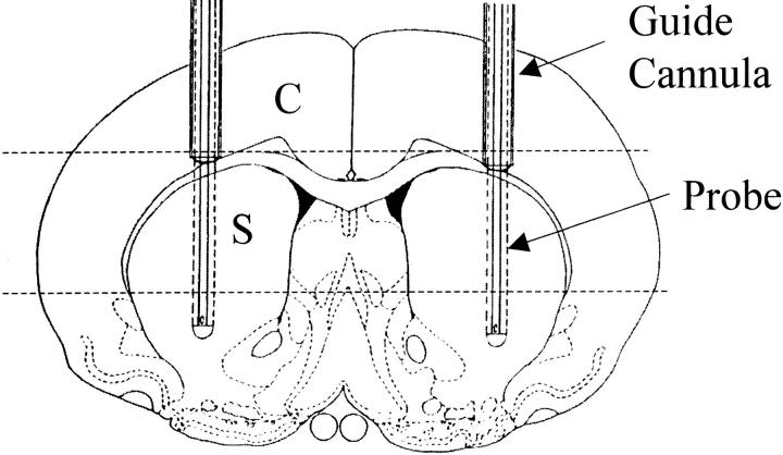
Figure 1.
Diagram of coronal view through striatum shows position of guide cannulae (outer diameter = 0.65 mm) and microdialysis probes (outer diameter = 0.5 mm with 4-mm-long dialyzing membranes). After fixation, brains were trimmed perpendicular to the probe tracks as indicated by dashed lines. Sections for histopathological analysis (6 μm) were taken through cortex (C) and striatum (S), perpendicular to guide cannulae and microdialysis probe tracks.

