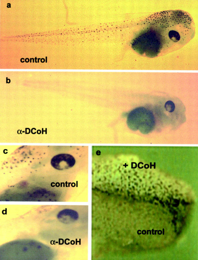
Figure 1.

Inhibition of DCoH/PCD during Xenopus embryogenesis suppresses pigmentation. a: Three-day old embryo (swimming larva, stage 42) with normal skin pigmentation. b: Larva of the corresponding stage derived from a fertilized egg that received DCoH/PCD-specific antibodies failed to develop dermal pigment cells. c and d: Eye regions of both larvae demonstrating that the ocular pigmentation is also disturbed (original magnification, ×4). Note some diffuse pigmentation below the eye in both larvae. e: Anterior region of a stage 25 embryo (2-day-old embryo) derived from a two-cell stage that was injected with DCoH/PCD mRNA into one blastomere (original magnification, ×8). Ectopic pigment cells were evenly distributed on the left side of the body, which expressed the introduced DCoH/PCD mRNA, whereas no dermal pigmentation is normally seen at this stage of development (right side).
