Figure 3.

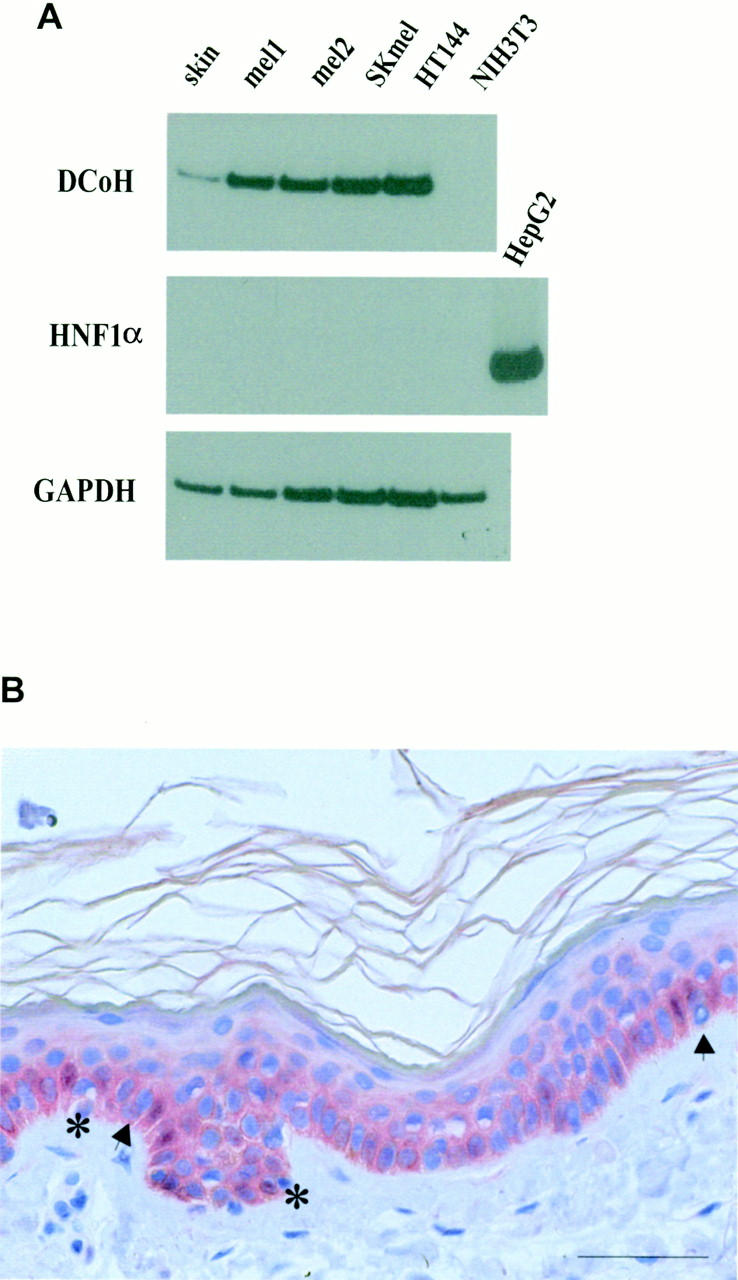
DCoH/PCD is detectable in primary human malignant melanoma lesions, melanoma cell lines, and normal human skin. A: RNA of different sources (lane 1, normal skin; lanes 2 and 3, two primary nodular melanoma lesions; lane 4, SK melanoma cell line; lane 5, HT144 melanoma cell line; and lane 6, NIH3T3 fibroblasts) was isolated and analyzed by RT-PCR for the presence of transcripts for DCoH/PCD (top), HNF1α (middle), and the housekeeping gene GAPDH (bottom). Control reactions performed without reverse transcriptase revealed no amplification (data not shown). B: Normal human skin immunostained with DCoH/PCD antibodies revealed faint staining of basal keratinocytes and melanocytes (arrows) (original magnification, ×200). Note the staining of the cytoplasm and the nucleus (asterisks) of individual cells. The immunoreactivity disappeared after preincubation of the antibodies with recombinant DCoH/PCD-protein (data not shown). Scale bar, 50 μm.
