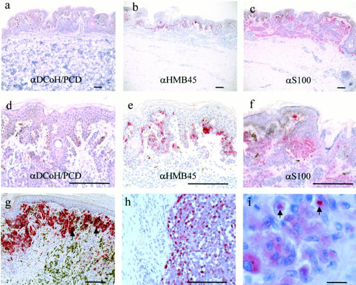
Figure 4.

DCoH/PCD is frequently overexpressed in primary melanoma lesions, but not in benign nevi. a: Benign nevi showed absent to weak DCoH/PCD staining in clusters of nevus cells (original magnification, ×50). For comparison, the staining of serial sections with anti-HMB45 (b and e) and anti-S100 (c and f) is also shown. d: Higher power view revealed the negativity of nevus cells for DCoH/PCD, whereas anti-HMB45 (e) and anti-S100 (f) stained nevus cells (original magnification, × 200). g: Primary superficially spreading melanoma lesions staining positive with DCoH/PCD polyclonal antibodies (original magnification, ×100). Note also some DCoH/PCD-positive melanophages (arrows). h: Higher power view of a different area of a primary nodular melanoma lesion demonstrates the characteristics of the DCoH/PCD staining with individual cells staining primarily in the cytoplasm (original magnification, ×200, respectively). Scale bars, 50 μm (a–h). i: Several melanoma cells show nuclear DCoH/PCD staining (arrows) (original magnification, ×1000). Scale bar, 50 μm (i).
