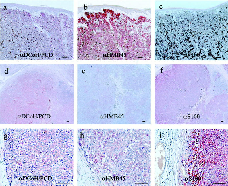
Figure 5.

DCoH/PCD expression in melanoma lesions is distinct from the melanoma markers S100 and HMB45. Serial sections of a primary superficially spreading melanoma lesion (a–c) and a primary nodular melanoma lesion (d–i) were either stained with the polyclonal antibodies specific for DCoH/PCD (a, d, and g), or commercially available monoclonal antibodies against HMB45 (b, e, and h), and S100 (c, f, and i). Original magnifications: ×100 (a–c); ×50 (d–f); and ×200 (g–i). Note the distinct staining pattern including cells that are negative for S100 and HMB45. Scale bars, 50 μm.
