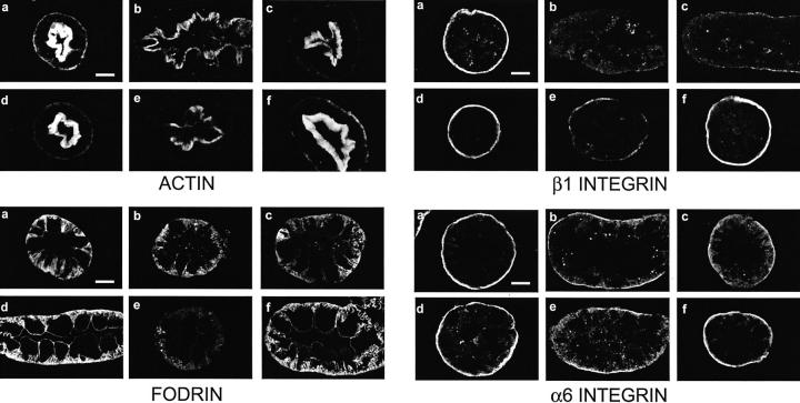
Figure 3.
Cytoskeletal alterations during hypoxia/reoxygenation. Tubules were subjected to 60 minutes of hypoxic incubation followed by 60 minutes of reoxygenation without or with supplemental αKG/ASP only during reoxygenation. ATP was added to the medium during reoxygenation. Samples fixed in paraformaldehyde-lysine-periodate at the end of hypoxia or after hypoxia and reoxygenation were cryosectioned and stained for F-actin with rhodamine phalloidin or immunostained for fodrin, or the β1 or α6 integrin subunits. In each of the sets of images, a and d are oxygenated time controls corresponding to the ends of the 60-minute hypoxic and 60-minute hypoxia plus 60 minutes reoxygenation periods, respectively. b and c are representative of the range of changes seen in tubules at the end of the 60-minute hypoxic period. The e panels show representative tubules after hypoxia followed by reoxygenation with NES. The f panels are from the paired flasks that were also subjected to hypoxia and reoxygenation, but were supplemented with αKG/ASP during reoxygenation. Magnifications are the same in each panel; scale bars, 10 μm.

