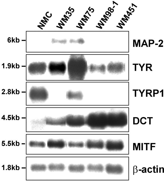
Figure 3.
Expression of MAP-2 mRNA in human melanocytes and melanoma cells. Northern blot analysis of 3 μg of polyA+ RNA isolated from cultured neonatal melanocytes (NMC), primary radial growth phase melanoma cell line WM35, primary VGP melanoma cell lines WM75 and WM98–1, and metastatic melanoma cell line WM451. Expression of MAP-2 and the melanocytic markers tyrosinase, TYRP1, DCT, and MITF were analyzed using 32P-labeled cDNA probes as described in Materials and Methods. Human β-actin probe was used to determine variation in RNA loading.

