Figure 6.

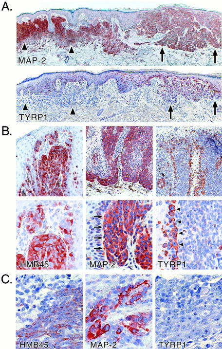
Immunohistochemical analysis of MAP-2 expression in melanocytic lesions. A: Primary malignant melanoma (superficial spreading type, Breslow thickness, 0.5 mm; Clark level III) arising in association with a melanocytic nevus. Upper panel: MAP-2. Lower panel: TYRP1. Arrowheads demarcate the approximate boundaries of the melanocytic nevus and the arrows indicate the melanoma. At this stage, the invasive melanoma cells are decorated as intensely as the nevus cells for MAP-2. Staining for TYRP1 is strong in the in situ melanoma but weak in the invasive disease and negative in the intradermal nevus cells. B: Primary malignant melanoma (Breslow thickness, 1.14 mm; Clark level III). Staining of invasive melanoma cells with anti-MAP-2 mAb is indicated by arrows and TYRP1 staining of tumor cells at the junction of the in situ and dermal invasive components of the tumor is highlighted by arrowheads. Note the reciprocal expression of TYRP1 and MAP-2. HMB45 staining helps to confirm melanocytic differentiation. Metastases of this melanoma, one excised from the left arm and another from the brain, stained negative or weakly positive for MAP-2, respectively (data not shown). C: The reciprocal relation between expression of MAP-2 and TYRP-1 is also illustrated in metastatic melanoma. HMB45 staining confirmed melanocytic differentiation of the metastatic lesion.
