
Figure 2.
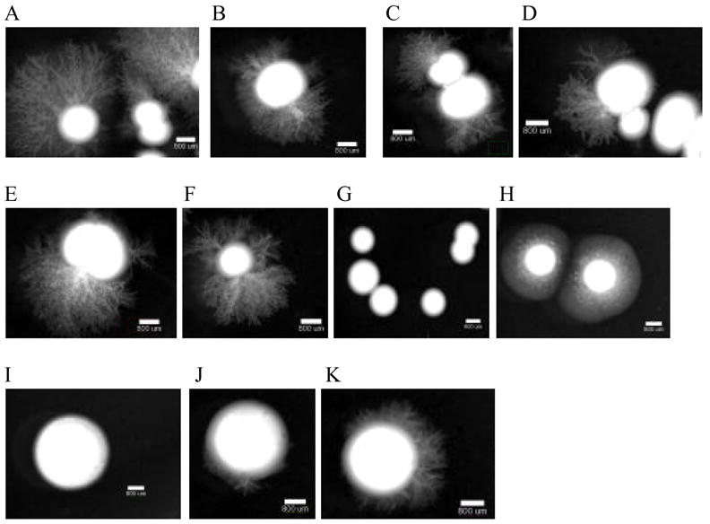
Filamentation assay on M199 pH 8. Each strain was grown overnight at 30°C in YPD liquid media. The cultures were diluted to a concentration of 10 cfu/μL, where 5 μL was added to medium 199 (M199) containing Earle’s salts and glutamine but lacking sodium bicarbonate (Gibco BRL) and buffered with 155 mM Tris-HCl at pH 8 + 1.5% agar. Pictures were taken at 14 day PI (A) SC5314, (B) DAY286, (C) ker1−/− (D) int1−/−, (E) muc1−/−, (F) mlt1−/−, (G) rim13−/−, (H) mds3−/−, (I) sla2−/−, (J) sch9−/−, and (K) suv3−/−. A and B are controls, C to F are mutants involved in adhesion or encode membrane proteins, and G to K are morphogenesis mutants.