
Fig. 4.
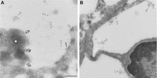
Cryo-immuno electron microscopic identification of GM3 ganglioside. Panel A: GM3 ganglioside is identified by 10 nm gold particles (arrowheads) whereas SCMAS is identified by 5 nm gold particles (arrows) and by the characteristic fingerprint-like array (asterisk) in lysosome of a pyramidal neuron. Panel B: GM3 is identified by 10 nm gold particles (arrowheads) in lysosome of a microglial cell. Scale bars are 0.2 μm.