
Figure 6.
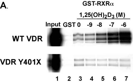
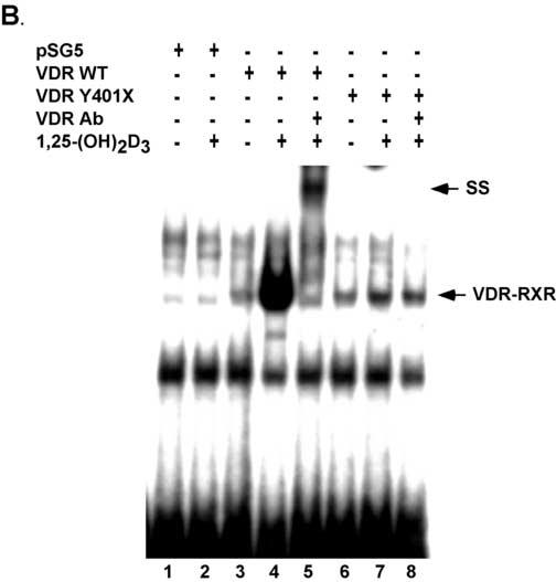
The truncated VDR binds RXRα and to VDREs. Panel A. VDRs labeled with [35S]-methionine by in vitro coupled transcription-translation were incubated with GST-RXRα in the presence of vehicle or graded concentrations of calcitriol. The samples were then subjected to GST-pull down assays and SDS-PAGE. Bands were visualized by autoradiography. The autoradiograph shows that the truncated VDR binds weakly to GST-RXRα and does not exhibit a 1,25(OH)2D3 dependent increase in binding as exhibited by the WT VDR. Panel B. Empty vector (pSG5) and WT and Y401X VDRs were expressed in COS-7 cells. Cell extracts were incubated with [32P]-labeled osteopontin VDRE with and without 10 nM 1,25(OH)2D3. For supershift assays VDR antibody was added. SS, supershift.