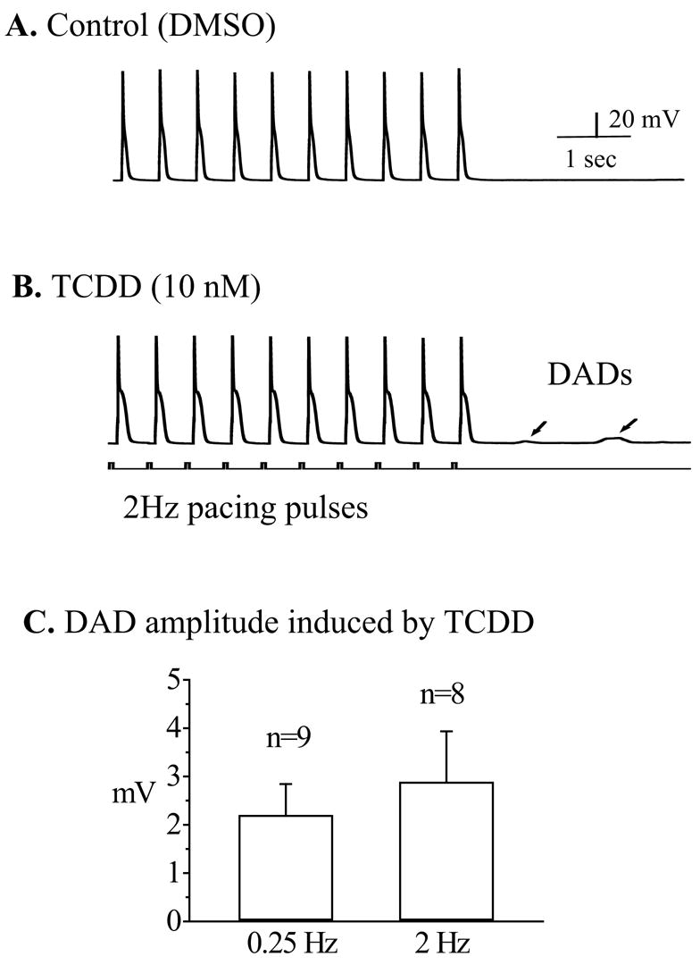
Figure 2.

TCDD induced delayed-afterdepolarizations (DADs). A train of action potentials (30 in total) was elicited by a 15 sec overdriving stimulation at a frequency of 2 Hz. Only last 10 action potential traces were shown. Recordings were carried out after either DMSO or TCDD (10 nM) application. A. A typical response of a ventricular mycyte treated with DMSO at 2 Hz pacing. Note that there was no after-depolarization activity when the pacing was terminated. B. In another myocyte treated with TCDD (10 nM), DADs (as indicated by arrows) were generated by the same stimulation protocol. Calibration marks apply to both panel A and B. Note, if there were multiple DADs in a single recording, the largest DAD was chosen to measure the amplitude of the DADs for the given cell.
