
Figure 3.
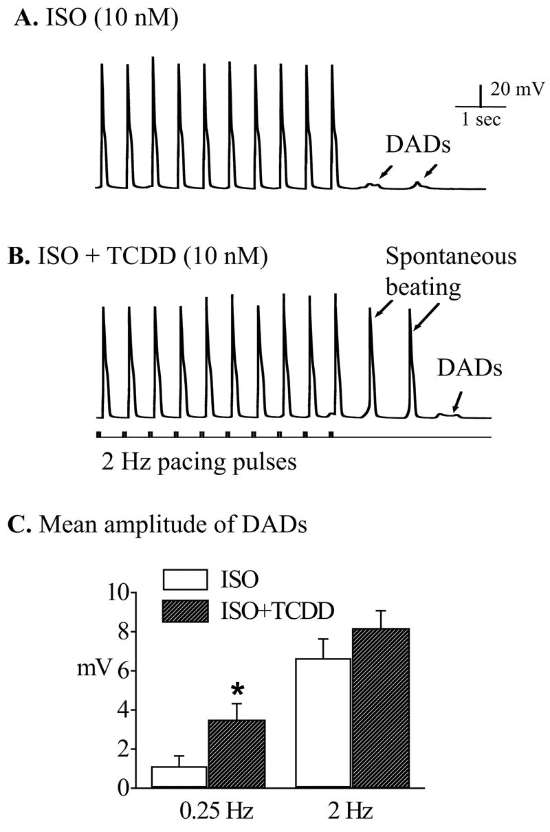
TCDD promoted ISO-induced DADs into spontaneous action potentials. Recordings were carried out after treating the cells with ISO (10 nM) alone or TCDD (10 nM) plus ISO (10 nM). A. A typical response of a ventricular mycyte treated with ISO (10 nM) to 2 Hz pacing. Note that there were after-depolarization activities when the pacing was terminated. B. In another myocyte treated with TCDD (10 nM) plus ISO (10 nM), DADs (as indicated by arrows) were developed into conductible spontaneous action potentials by the same stimulation protocol. Calibration marks apply to both A and B. C. The average amplitudes of DADs induced by ISO alone and ISO plus TCDD at two pacing frequencies (0.25 and 2 Hz). * indicates statistic significance at p<0.05 level.