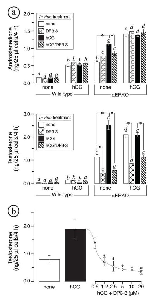
FIG. 5.

In vitro evaluation of HSD17B3 activity in dispersed αERKO ovarian cells. All data are from in vitro acute steroidogenic assays on dispersed ovarian cells (see Materials and Methods). These experiments were repeated three times and found to yield comparable results, shown are the results of one independent trial. Wild type (WT) and αERKO females were first treated twice daily for 3 days with vehicle or human choriogonadotropin (hCG) as indicated along the x-axis; dispersed ovarian cells were then prepared from each genotype/treatment group and incubated for 4 h in the indicated in vitro treatments. (a) In vitro androstenedione (top) and testosterone (bottom) synthesis indicates that in vivo hCG treatments led to increased androstenedione production in both WT and αERKO ovarian cells but increased testosterone synthesis in αERKO cells only. In vitro testosterone synthesis in dispersed αERKO ovarian cells was inhibited by the HSD17B3 inhibitor (DP3-3) at 10 μM. (b) Shown is a dose-response curve illustrating the effect of the HSD17B3 inhibitor (DP3-3) on testosterone synthesis in dispersed αERKO ovarian cells. All data are from dispersed ovarian cells from untreated αERKO females that were incubated in medium alone, medium with hCG (10 I.U./ml), or medium with hCG (10 I.U./ml) plus increasing concentrations of DP3-3. Each bar or point represents the average of 3 replicates, each of which was assayed in duplicate for each steroid. Bars that do not share the same letter are significantly different (P < 0.05). Significant differences (P < 0.05) within each genotype/treatment group are indicated by asterisks.
