
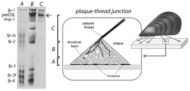
FIGURE 1.
Schematic drawing of the mussel byssus illustrating a close-up of the junction between the thread and plaque portions and the extensive interface between the splayed collagen fibrils and foam proteins of the plaque (right). Gel electrophoresis (acid urea-PAGE) of the proteins extracted from three microdissected portions of freshly secreted byssal threads (left) [(A) footprint region, (B) junction, and (C) distal thread]. The gels have been stained for Dopa with glycinate and nitroblue terazolium (NBT). Identification of known protein families is according to previous studies (9-16). The boxed band is mcfp-4, and the double-headed arrow marks the position of an internal calibrant, the α chain of type I collagen.