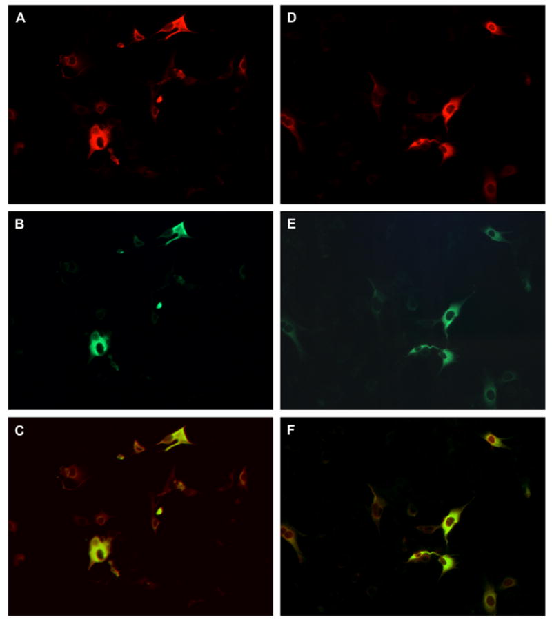
Fig. 4.

Double-staining of BHK transfectant expressing mEH. After transfection, the cells were transferred to a chamber slide and cultured. Then they were fixed with acetone and double-stained by rabbit anti-mEH and mouse anti-AN mAb 1F12 (A and B), or by rabbit anti-AN antibody and 1F12 (D and E). Rabbit and mouse antibodies were detected by TRITC- and FITC-labeled second antibodies, respectively. The merged image of A and B and that of D and E are shown in C and F, respectively.
