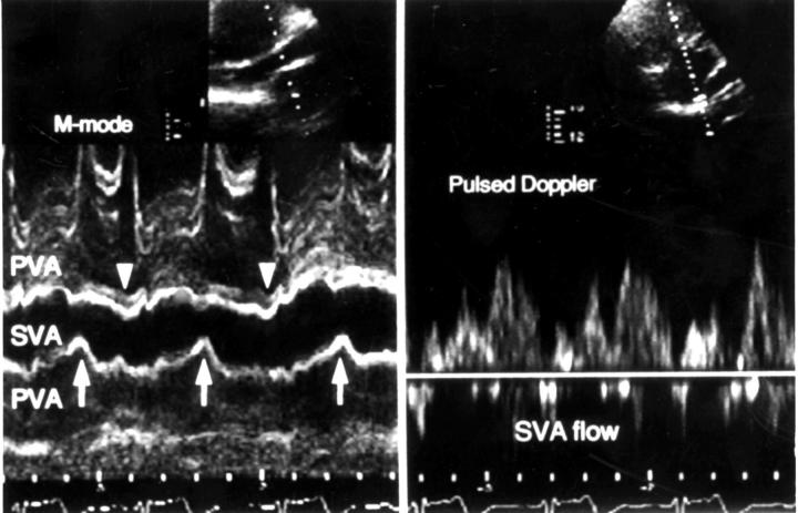
Figure 4 .
M mode (left panel) and pulsed Doppler (right panel) findings after Senning procedure. In mid-diastole (arrows), the systemic venous atrium (SVA) is compressed by pressure from surrounding pulmonary venous atrium (PVA). This causes a decrease in left ventricular filling rate until the SVA reopens due to a pressure increase proximal to the narrowed segment. The filling rate then increases again, with only a small contribution from atrial contraction (arrowhead). See Discussion.

